
The skin is something we often take for granted—until something goes wrong. Whether it’s dryness, irritation, loss of elasticity, or visible aging, the condition of our skin quickly becomes a reflection of both our internal health and our external habits. But beyond its appearance, the skin is a highly sophisticated, dynamic organ performing essential life-supporting functions every second.
Understanding how the skin is structured—and what happens within its layers—can completely change the way we approach skincare. Instead of relying on trends or guesswork, we begin to make informed choices that truly support the skin’s natural processes. This knowledge allows us to work with the skin rather than against it.
In this article, we’ll explore the anatomy and physiology of the skin, break down its layers, and take a closer look at the key components that determine its strength, elasticity, hydration, and overall health.
The skin is our largest organ, accounting for about 15% of total body weight. Its surface area is approximately 2 square meters, and it makes up roughly one-sixth of a person’s total weight. It may not be the largest in size compared to internal organs, but it is certainly the most visible. That is why the condition of the skin has such a strong impact on self-esteem.
The skin is highly sensitive: it can detect both the lightest touch and pain. One square inch (2.5 cm) of skin contains about 19 million skin cells and 60,000 melanocytes (cells responsible for producing skin pigment). Every 2.5 cm of skin also contains around 1,000 nerve endings and 20 blood vessels.
Our skin performs many functions. First and foremost, it protects the body from external physical, chemical, and biological factors, prevents water loss, and participates in thermoregulation. Recent scientific findings confirm that the skin not only has its own immune system but also functions as a peripheral immune organ.
It serves as the first line of defense against bacteria and viruses while maintaining the body’s water balance. The health of the skin largely determines proper thermoregulation, protection against pathogens, and, of course, physical appearance.
The skin is vital for our health and overall well-being. To care for it properly across all areas, it is important to understand how it works—its structure, composition, and functions.
Anatomy and Physiology of the Skin
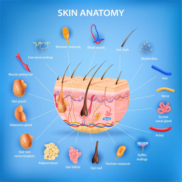
Our skin consists of several layers, each with its own sublayers. The structure of human skin includes:
• the outer layer (epidermis);
• the middle layer (dermis, or corium);
• the inner layer, or subcutaneous fat tissue (hypodermis).
Skin appendages—hair follicles, sebaceous glands, and sweat glands—also help the skin perform its functions.
Epidermis
This is the outer layer of the skin—the part we see and touch. The epidermis protects us from toxic substances, harmful microorganisms, and fluid loss. It is the uppermost and thinnest layer of the skin and plays a key role in protecting the body from external influences.
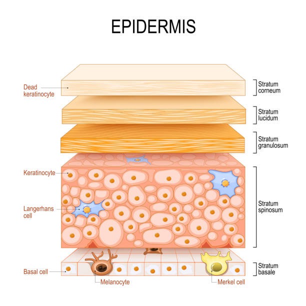
The epidermis is divided into five layers composed of keratinocyte cells: the stratum corneum, stratum lucidum, stratum granulosum, stratum spinosum, and stratum basale. These layers differ in the stages of keratinocyte maturation.
1 Stratum basale (basal layer): the deepest layer. Here, keratinocytes divide, begin their development, and gradually move toward the skin’s surface. As they migrate, they mature and change—a process known as keratinization. This layer also contains immune cells and melanocytes. Melanin provides protection from UV radiation and determines skin color.
2 Stratum spinosum (spinous layer): composed of cells connected by desmosomes, which give the epidermis strength. Here, keratinocytes produce keratin (protein fibers) and take on a spindle-like shape.
3 Stratum granulosum (granular layer): in this layer, cells begin producing keratohyalin, a precursor of keratin. Keratinization starts here—cells produce dense granules that gradually transform into keratin and epidermal lipids as they move toward the surface.
4 Stratum lucidum (clear layer): cells become flattened and tightly packed, and their boundaries become less distinct. This layer is present only in areas of thick skin, such as the palms and soles, providing additional strength. It is absent on areas like the face and neck.
5 Stratum corneum (horny layer): the outermost layer of the epidermis, consisting on average of about 20 sublayers of flat, dead keratinocytes that continuously shed and renew. Openings of sweat and sebaceous gland ducts are also located here.
 Any product we apply to the skin primarily acts within the epidermal layer, and only a small portion of active ingredients penetrates deeper.
In a healthy state, the epidermis has a balanced microflora (a community of beneficial bacteria) that prevents the penetration of harmful substances (pathogens) and contributes to skin immunity. The epidermis also contains a natural protective barrier known as the hydrolipid mantle. It is composed of corneocytes (dead skin cells), water molecules, and lipids. This barrier helps protect against external aggressors and prevents transepidermal water loss.
The epidermis constantly produces new skin cells, replacing approximately 40,000 old cells shed by the body every day. Complete skin renewal typically takes about 30 days. With proper care — combining timely removal of dead cells, restoration of natural resources, and support of the protective barrier — it is possible to achieve radiant, healthy-looking skin.
To support and accelerate this process, the use of physical and chemical exfoliants is recommended: peels, scrubs, enzyme powders, and similar products.
Dermis (Corium)
The dermis is the second layer of the skin, characterized by a thicker structure and rich blood supply. It is a complex, loosely organized connective tissue composed of fibers, cells, a network of blood vessels and nerve endings, as well as epidermal projections surrounding hair follicles and sebaceous glands.
The cellular components of the dermis include fibroblasts, macrophages, and mast cells. Lymphocytes, leukocytes, and other cells can migrate into the dermis in response to various stimuli. Compared to the epidermis, the dermis is significantly thicker—normally up to 5–6 mm—and is both elastic and dense.
It consists of two main layers:
1 Papillary layer (stratum papillare): the upper part of the dermis, located directly beneath the epidermis. It is made of loose connective tissue and contains capillaries, nerve endings, and small projections called papillae. This layer supplies nutrients to the epidermis and contributes to thermoregulation and skin sensitivity.
2 Reticular layer (stratum reticulare): the deeper and thicker part of the dermis, forming a flexible boundary with the hypodermis. It contains collagen and elastin fibers, which provide strength and elasticity. It also houses blood vessels, nerves, hair follicles, and sebaceous and sweat glands.
Blood vessels in the dermis supply oxygen and nutrients to the epidermis, supporting its regeneration and proper functioning. They also regulate heat exchange, helping the body maintain an optimal temperature.
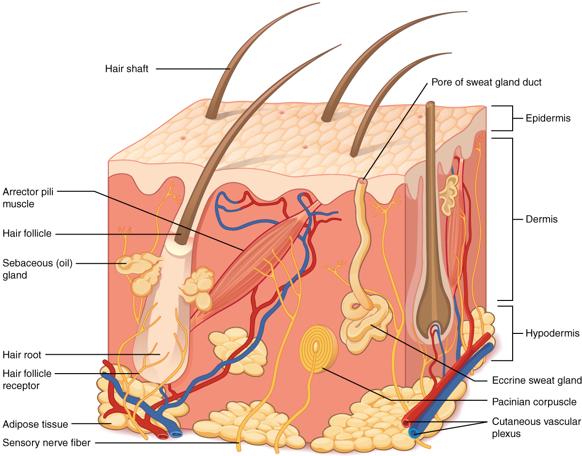
The dermis acts as a supporting structure for the epidermis, providing attachment and stability. Its structure includes fibroblasts, collagen and elastin fibers, and an intercellular matrix.
Sebaceous glands (which secrete sebum) and sweat glands (which release water and lactic acid) are located in the dermis. Their secretions combine to form the hydrolipid film.
The dermis also contains:
- Lymphatic vessels
- Sensory receptors
- Hair follicles
The main structural components of the dermis are collagen and elastin—proteins that provide strength and elasticity. They are essential for maintaining healthy, youthful skin. These fibers are surrounded by a gel-like substance (the extracellular matrix), which contains hyaluronic acid. This substance can retain large amounts of water and helps maintain skin volume.
Lifestyle and environmental factors, such as sun exposure and temperature fluctuations, have a destructive effect on collagen, elastin, and the extracellular matrix. As we age, the natural production of collagen and elastin slows down, and the skin’s ability to retain moisture decreases. As a result, the skin loses tone and wrinkles appear.
The dermis is the layer where most processes affecting skin firmness and youthfulness occur. Its structure includes collagen and elastin, while the space between them is filled with the extracellular matrix — a gel-like substance composed mainly of water and hyaluronic acid.
Blood vessels, lymphatic vessels, and nerve endings are also present in the dermis. However, the most important structural elements are fibroblasts—cells that produce collagen, elastin, and hyaluronic acid. The firmness of the skin largely depends on the condition of these fibers and the activity of fibroblasts. With age, their activity gradually decreases, causing the dermal layer to thin and lose its density, leading to reduced elasticity.
Proper dermal care includes hydration, stimulation of collagen production, and protection from environmental damage. Products containing hyaluronic acid of different molecular sizes, peptides that influence fibroblast activity, and vitamins are particularly beneficial.
Subcutaneous Fat Layer (Hypodermis)
The subcutaneous fat layer, or hypodermis, is the deepest layer of the skin, located beneath the dermis. While smaller blood vessels are found in the dermis, larger arteries, veins, and nerves run through the hypodermis.
It consists of fat lobules separated by connective tissue septa containing collagen and penetrated by larger blood vessels. The main cells of these fat lobules are adipocytes, whose number varies across different parts of the body.
The hypodermis cushions impacts and helps retain body heat. Its primary function is to store energy in the form of fat, which can be released when needed, such as during physical activity or nutrient deficiency.
The structure of the hypodermis varies across different areas of the body. On the face and hands, it may be relatively thin, while in areas such as the abdomen or buttocks, it is thicker due to a higher number of fat cells. Fat distribution also differs between men and women.
The hypodermis mainly consists of:
- Fat cells (adipocytes) grouped into dense clusters
- Connective tissue that holds these cells in place
- Blood vessels supplying nutrients
- Nerve endings transmitting sensory signals
The hypodermis plays an important role in thermoregulation. Fat cells generate heat and act as insulation, helping the body maintain a stable internal temperature.
Today, the hypodermis is considered not only an energy reserve but also an endocrine organ. Its cells produce hormones and signaling molecules that influence metabolism, insulin sensitivity, and immune function.

A Brief Overview of Key Components in Skin Structure
The extracellular matrix (ECM) of the dermis, or intercellular substance, consists of various proteins (primarily collagen and elastin), glycosaminoglycans—most notably hyaluronic acid—and proteoglycans such as fibronectin, laminin, decorin, versican, and fibrillin. All of these substances are produced by dermal fibroblasts.
The ECM is not a random collection of elements but a highly organized network. Its structure determines the biomechanical properties of the skin, including firmness, elasticity, and flexibility. Keratinocytes of the epidermis attach to ECM proteins and form a tightly connected protective layer.
Fibroblasts are the main cellular components of the dermis. They are responsible for producing collagen, elastin, hyaluronic acid, and other essential proteins. Their activity varies depending on their depth within the dermis and external stimuli. These cells are highly adaptable and can change their function in response to environmental signals.
Collagen is the primary structural protein of the dermis, making up about 70% of its dry weight. It provides strength and support, forming the framework of the skin. Collagen is constantly renewed, but its production declines with age, leading to loss of firmness and the appearance of wrinkles.
Elastin forms a flexible network that allows the skin to stretch and return to its original shape. Without sufficient elastin, the skin loses its resilience and smoothness.
Hyaluronic acid plays a crucial role in maintaining hydration. It binds water molecules and supports the structure of the extracellular matrix, contributing to skin volume and firmness. It also influences cell movement and immune responses within the skin.
Vitamins
Vitamin A promotes cell renewal, vitamin C supports collagen production, and vitamin E protects the skin as a powerful antioxidant. Including these vitamins in skincare helps maintain healthy, youthful-looking skin.
Antioxidants
Antioxidants protect the skin from oxidative stress caused by free radicals. Ingredients such as coenzyme Q10, green tea, and vitamin C help prevent premature aging, reduce damage, and support overall skin health.
The skin is far more than just a surface—it is a complex, living system where structure and function are deeply interconnected. Each layer, from the protective epidermis to the supportive dermis and the insulating hypodermis, plays a unique and essential role. Within these layers, key components such as collagen, elastin, hyaluronic acid, and fibroblasts work together to maintain strength, elasticity, hydration, and resilience.
When we understand how these elements interact, skincare becomes more intentional and effective. Instead of focusing only on surface-level results, we begin to support the deeper processes that truly determine the condition of the skin.
Healthy skin is not achieved through a single product or quick fix—it is the result of consistent care, informed choices, and respect for the skin’s natural biology. By learning how the skin functions and what it needs, you gain the ability to care for it in a way that enhances both its appearance and its long-term health.